The eye is a window to the retinal vascular system which is uniquely accessible for the non-invasive, in vivo study of a continuous vascular bed in humans. The detection and measurement of blood vessels can be used to quantify the severity of disease, as part of the process of automated diagnosis of disease or in the assessment of the progression of therapy. Retinal blood vessels have been shown to have measurable changes in diameter, branching angles, length or tortuosity, as a result of a disease [1,2,3]. Thus a reliable method of vessel segmentation would be valuable for the early detection and characterisation of changes due to such diseases.
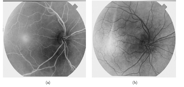
Different techniques are used to acquire images of retinal blood vessels. A relatively non-invasive technique, widely used clinically, is the retinal fundus photograph taken using a green filter, generally called a red-free image. A more invasive technique is fluorescein angiography which involves an intravenous injection of dye which increases the contrast of the blood vessels against the background. Figure 1 shows an example of two scanned negatives taken from the same eye before (red-free) and after (fluorescein) the injection of fluorescein dye.
There have been many studies on the detection of blood vessels in medical images
in general but only a small number are related to retinal blood vessels in
particular. Most of the work on segmentation of retinal images can be categorised
into two approaches: those based on line or edge detectors with boundary tracing
[4,5] and those based on matched filters, either 1-D profile matching
with vessel tracking [6,7,8,9] or 2-D matched filters
[10,11,12].
We have applied some of these methods but
because of the large regional variations in intensity inherent in these images
and the very low contrast between vessels and the background, particularly in the
red-free photographs, the results were disappointing. Techniques based on line or
edge detectors lacked robustness in defining blood
vessels without fragmentation and techniques based on matched filters
were difficult to adapt
to the variations of widths and orientation of blood vessels.
Furthermore all of these methods are developed to work either on
red-free or fluorescein images but not on both.
In this paper we present a method based on multiscale
analysis from which we obtain retinal blood vessel width approximation, size and
orientation using gradient
magnitude and maximum principal curvature of the Hessian tensor, two geometrical features
based upon the
first and the second spatial derivatives of the intensity considered along the scales
that give information about the topology of the image at different scales.
We then use a multiple pass
region growing procedure which progressively segments the blood
vessels using the feature information together with spatial
information about the 8-neighbouring pixels, obtaining in this way
a segmented binary image. The algorithm works equally well with both
red-free fundus images and fluorescein angiographs.
![]()
![]()
![]()
Next: The Segmentation Method
Up: Retinal Blood Vessel Segmentation
Previous: Retinal Blood Vessel Segmentation