

Next: Validation and Comparison Between
Up: Retinal Blood Vessel Segmentation
Previous: Region growing
The retinal photographs tested were taken using a fundal camera with a  field of view (Kowa FX-50R, Kowa, Tokyo, Japan) using Ilford FP4 (125 ASA) photographic
film (Ilford Imaging UK Ltd., Knutsford, England). Photographic
negatives were digitised using a Nikon 35
  film scanner (LS-1000, Nikon, Tokyo, Japan).
Digitised images were
 pixels in size.
For the analysis of complete fundus images, these were reduced to
600 x 700
pixels by applying a Gaussian pyramid low-pass filter [23].
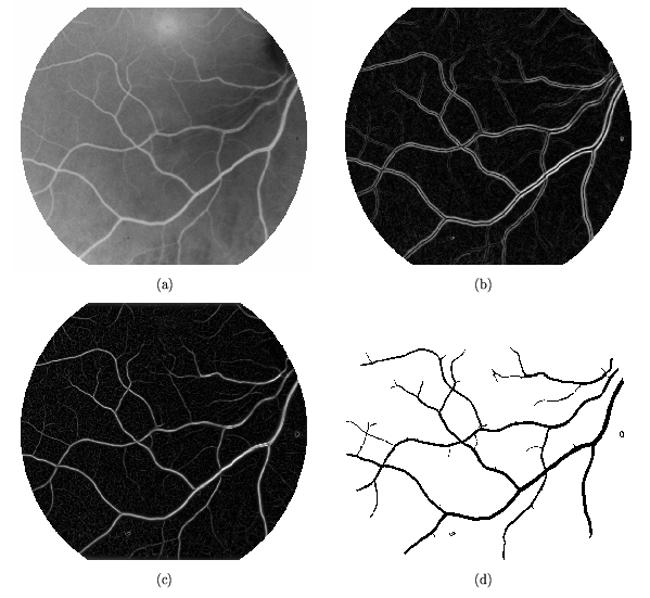
Figure 10 shows the segmentation process described in the previous
section, (a) negative red-free gray scale image, (b)  , (c)
, (c)  and (d) the segmented image. For the reduced size images,
an interval of
 pixels was used.
Figure 10:
The segmentation method. (a) Original, (b)  ,
(c)
,
(c)  and (d) segmented. Image size 600 x 700
pixels.
 |
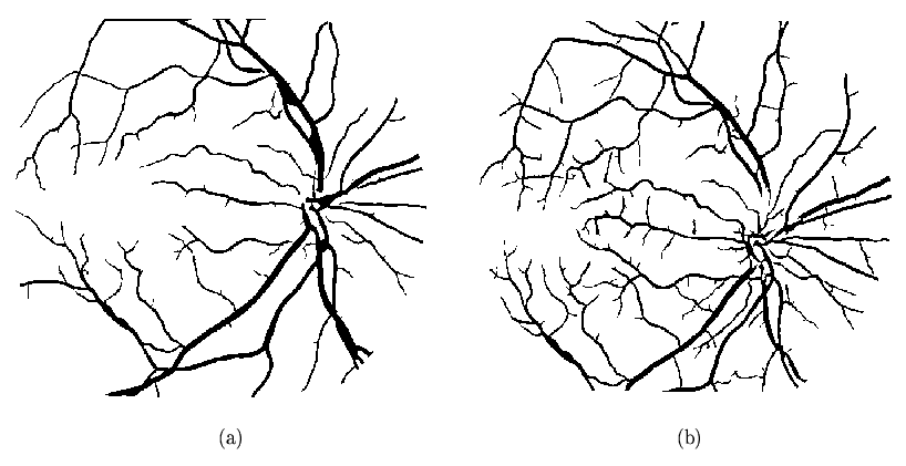
Figure 11 shows the segmented images corresponding to the original images
shown in Figure 1, red-free and fluorescein.
Figure 11:
Segmented images from Figure 1, respectively, (a) red-free,
(b) fluorescein. Image size 600 x 700
pixels.
 |
It can be seen that despite the much poorer contrast, the
red-free image segmentation is nearly as good as that of the fluorescein image. We
also note that the segmentation is relatively insensitive to the wide variations
in intensity that are inherent in these images. In the fluorescein image,
for example, the detail in the segmented image in the much brighter
region just beside the darker optic disk in the left centre of the image is
nearly the same as in the higher contrast portions of the image.


Next: Validation and Comparison Between
Up: Retinal Blood Vessel Segmentation
Previous: Region growing
Elena Martínez
2003-05-16
![]() field of view (Kowa FX-50R, Kowa, Tokyo, Japan) using Ilford FP4 (125 ASA) photographic
film (Ilford Imaging UK Ltd., Knutsford, England). Photographic
negatives were digitised using a Nikon 35
field of view (Kowa FX-50R, Kowa, Tokyo, Japan) using Ilford FP4 (125 ASA) photographic
film (Ilford Imaging UK Ltd., Knutsford, England). Photographic
negatives were digitised using a Nikon 35 ![]() film scanner (LS-1000, Nikon, Tokyo, Japan).
Digitised images were
film scanner (LS-1000, Nikon, Tokyo, Japan).
Digitised images were
![]() pixels in size.
For the analysis of complete fundus images, these were reduced to
600 x 700
pixels by applying a Gaussian pyramid low-pass filter [23].
pixels in size.
For the analysis of complete fundus images, these were reduced to
600 x 700
pixels by applying a Gaussian pyramid low-pass filter [23].
![]() , (c)
, (c) ![]() and (d) the segmented image. For the reduced size images,
an interval of
and (d) the segmented image. For the reduced size images,
an interval of
![]() pixels was used.
pixels was used.

